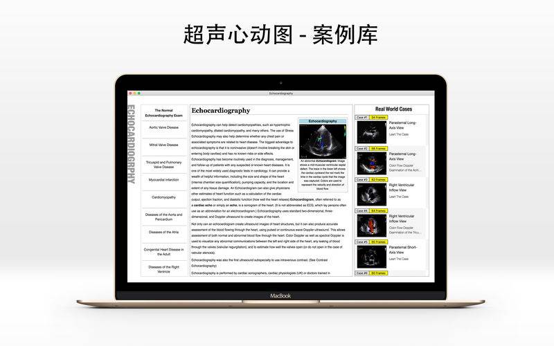
应用包含200多例心动图的完整病例,涵盖20多种与心血管系统有关的疾病,以及疾病在心动图上的表现和分析方法。此应用可将您的Mac电脑变成专业的心动图分析工作站。
超声心动图,是一种心脏超声波检查,它使用标准的超声波技术显示心脏的二维图片。现在最新的超声诊断系统采用三维实时成像。耗時大約15-20分鐘,甚至更長。
除了产生心血管系统的两维图像,超声心动图使用脉冲或者连续超声波,能对任意位置的血液和心肌组织速度作出准确的测量。这样可以检测心脏瓣膜区域功能、左右侧心脏不正常联系、瓣膜返流、以及心脏输出量的计算等。其他测量的参数包括心脏尺寸(管腔直径和室间隔厚度)和E / A比值。
Turn your Mac to an Echocardiography Workstation, with over 200 real world echocardiography exams to review and learn. Get yourself ready before you do the job on real human’s heart.
Echocardiography, often referred to as a cardiac echo or simply an echo, is a sonogram of the heart. (It is not abbreviated as ECG, which lay persons often use as an abbreviation for an electrocardiogram.) Echocardiography uses standard two-dimensional, three-dimensional, and Doppler ultrasound to create images of the heart.
Echocardiography has become routinely used in the diagnosis, management, and follow-up of patients with any suspected or known heart diseases. It is one of the most widely used diagnostic tests in cardiology. It can provide a wealth of helpful information, including the size and shape of the heart (internal chamber size quantification), pumping capacity, and the location and extent of any tissue damage. An Echocardiogram can also give physicians other estimates of heart function such as a calculation of the cardiac output, ejection fraction, and diastolic function (how well the heart relaxes).
Echocardiography can help detect cardiomyopathies, such as hypertrophic cardiomyopathy, dilated cardiomyopathy, and many others. The use of Stress Echocardiography may also help determine whether any chest pain or associated symptoms are related to heart disease. The biggest advantage to echocardiography is that it is noninvasive (doesn’t involve breaking the skin or entering body cavities) and has no known risks or side effects.
Not only can an echocardiogram create ultrasound images of heart structures, but it can also produce accurate assessment of the blood flowing through the heart, using pulsed or continuous wave Doppler ultrasound. This allows assessment of both normal and abnormal blood flow through the heart. Color Doppler as well as spectral Doppler is used to visualize any abnormal communications between the left and right side of the heart, any leaking of blood through the valves (valvular regurgitation), and to estimate how well the valves open (or do not open in the case of valvular stenosis).
Echocardiography was also the first ultrasound subspecialty to use intravenous contrast.